Cone Beam CT: An Interview with the President of iCat
We recently had the opportunity to interview Ed Marandola, the President and CEO of Imaging Sciences International, about their cutting-edge, 3-D, cone beam CT scanning technology, the i-CAT. Their facility is located in Hatfield, Pennsylvania, just outside of Philadelphia. Their website is www.imagingsciences.com and telephone number is 800-205-3570. 3-D Cone beam CT scanning represents the next major leap in new technology, and the Imaging Sciences’ i-CAT has become the leader.
Osseonews: What are the advantages of cone beam CT scanning over the traditional fan beam CT scans?
Ed Marandola: There are many advantages over traditional CT scans. First, with cone beam CT scanning the patient is subjected to far less radiation, because we use a focused beam with a physically larger sensor allowing us to capture all the axial slices covering the face and the jaws in one scan. Since we capture the entire volume with one scan, there is no redundant overlap of slices, resulting in major reduction of radiation. Reducing exposure to radiation while maximizing the information content should be a goal of all diagnostic imaging and we accomplish that very effectively with our i-CAT system.
The single most groundbreaking advantage of the i-CAT is easy, convenient and low-cost access to this imaging modality for the patient and the dentist. When this machine is in-office and the dentist has made the capital investment, superior and invaluable diagnostic information is produced for the patient whenever warranted, without the obstacles that forced compromises in the past.
Osseonews: How would you compare the information you generate with cone beam CT scanning to information generated by an MRI or a panoramic radiograph?
Ed Marandola: A panoramic radiographic provides a 2-dimensional depiction of the patient. All panoramic radiographs are distorted up to 300% in the horizontal plane and up to 50% in the vertical plane. A cone beam CT scan, such as our i-CAT, provides a true reproduction of the patient’s anatomy. The dimensions represented in the cone beam imaging correspond exactly to the patient. There is no distortion. The dimensions are accurate. MRI’s are more useful for soft tissue imaging.
Osseonews: What is the most efficient way to use your cone beam imaging for treatment planning for dental implants?
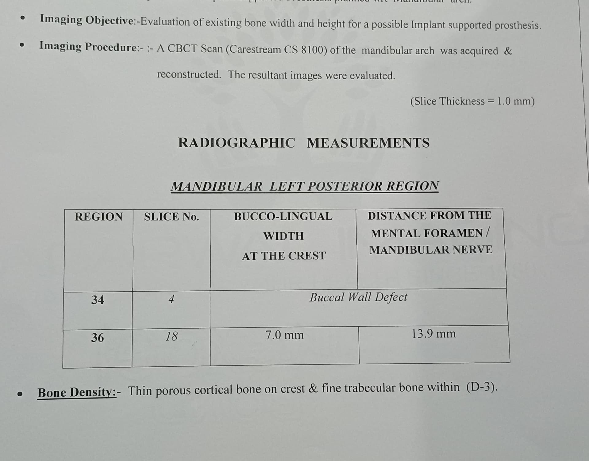
Ed Marandola: After scanning the patient the doctor can generate an exact reproduction of the patient’s mouth, face and jaw area. The doctor can identify the location and dimensions of all anatomic structures such as the maxillary sinus or inferior alveolar nerve or mental foramen nerve. The doctor can also measure the exact dimensions and quality of the bone, which is crucial for implant placing procedures.
The i-CAT(TM) Cone Beam 3-D Imaging System produces immediate three-dimensional images of patients’ critical anatomy, typically in under one minute. The i-CAT (TM) provides complete views of all oral and maxillofacial structures in an easy to use, cost effective, in-office system that allows dental specialists to dramatically enhance their patient care in a variety of ways.
The immediate three-dimensional reconstruction of a patient’s mouth, face, and jaw areas, with the i-CAT (TM) enhances doctor/patient communication by allowing doctors to share a visual diagnosis with their patients. This gives patients a better understanding of their treatment options and more confidence when going into treatment.
Osseonews: Can the imaging be used to plan for the exact placement of implants?
Ed Marandola: Our cone beam scanning reproduces the anatomy of the patient. The i-CAT software is compatible with other software programs from implant manufacturers and you can use their software with our imaging to plan with great accuracy and confidence which kind of implant to place. You can use their software to place implants of varying design, width and length into our i-CAT imaging to determine the most efficacious design, site, angulation and dimensions. There is no guesswork involved. What you plan is what you get.
Osseonews: Can you use your narrow cone imaging with subperiosteal implants?
Ed Marandola: We have been doing this very successfully. The doctor can use our cone beam CT scan to generate an exact reproduction of the maxilla or mandible. Our scan is transmitted to a dental laboratory that custom makes the metal subperiosteal framework and it fits exactly on the alveolar ridge. The advantage to using our cone beam CT scan for this is that it completely eliminates the need for surgical exposure and impressioning of the alveolar ridge.
Compared to medical scanners, Cone Beam Scanning is ten times more accurate while reducing a patient’s exposure to radiation by more than 95%. Pre-surgical implant treatment planning, preparing to remove impacted third molars, determining how sinus grafts and ridge augmentations have healed, and determining the ideal position for a single-tooth replacements, are just some of the benefits of Cone Beam Scanning Technology. Since Cone Beam Scanning permits multiple slices through the axial, sagittal and coronal views, the guesswork is removed when it is critical to determine the width of edentulous ridges, whether or not cancellous bone exists between cortical plates, the position of supernumerary and developing tooth buds, if sockets have filled with bones, if irregularities exist to the condyles, where the mandibular nerve is relative to an impacted tooth and implant sites, or to visualize the borders of a cyst or tumor. Cone Beam Scanning has an added benefit in that it can take the maxilla and mandible in a single scan.
Osseonews: How big is your cone beam unit?
Ed Marandola: The i-CAT is slightly wider than a panoramic radiograph. The patient remains seated as the i-CAT rotates around the head for a 20 second scan.
Osseonews: What is the cost for purchasing and installing the cone beam unit?
Ed Marandola: The total cost for purchase, installation and training is $155,000. Compare that to a full-service CT scanner which costs upwards of half a million dollars or more.
Osseonews: How long does it take to train the auxiliary staff to take cone beam CT scans?
Ed Marandola: It takes about a day. We come out and spend an entire day training the staff and having them practice.
Osseonews: What happens if they run into problems in the future?
Ed Marandola: We have a help desk that they can call. We can also link up with any of the CT units we install via the internet and we can then diagnose or trouble-shoot directly out of information in the unit.
Osseonews: What kind of feed-back have you been getting from doctors who have had the cone beam CT scans installed in their offices?
Ed Marandola: We hear comments like “We didn’t know what we didn’t know (about the patient’s anatomy)†and “Can’t live without itâ€. We constantly hear very positive comments and have very happy customers.
Interviewed provided by Dr. Gary Kaplowitz, Special Editor for OsseoNews.com


