Dull ache from implant: how to manage?
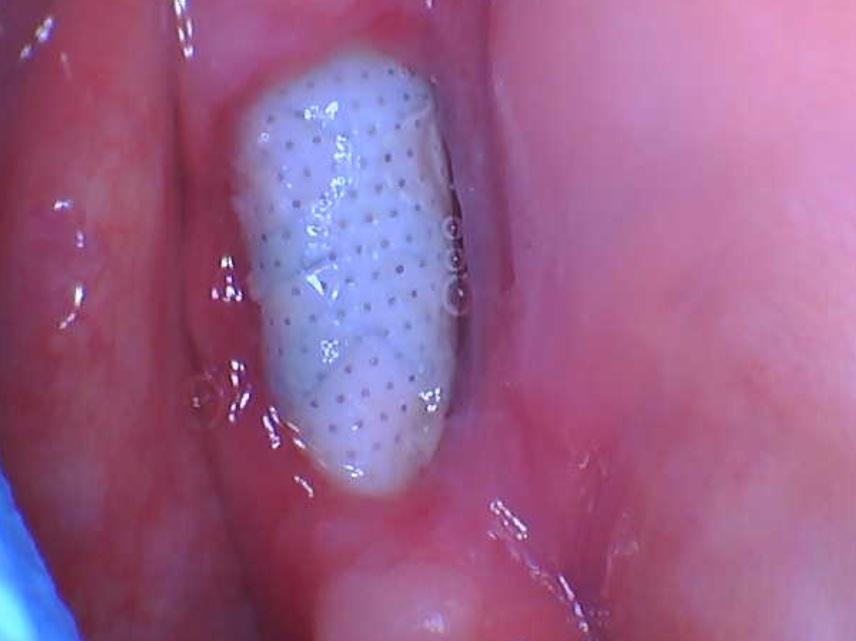
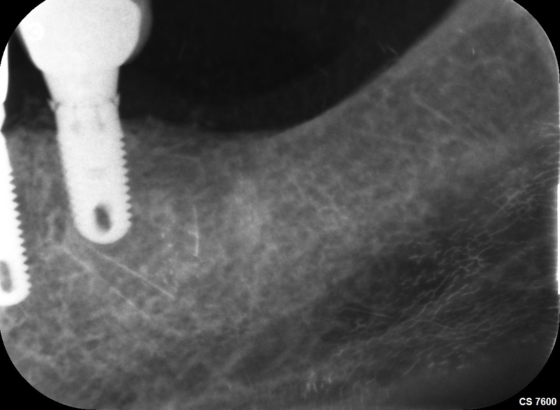
Patient is fit and healthy middle aged female. 1 year ago upper front anteriors were lost due to trauma and had implants placed 1 year ago in UR1 and UL1 to support a 4 unit upper anterior bridge. Clinically no mobility, not tender to percussion, soft tissues look excellent, probing depths around implants 3-4mm, no BOP or exudate. She is complaining of a dull ache in the area of 21 implant buccal gingivae base of sulcus, which is relieved when pressing on the gum where the implant apex would be. It is not a pain where it affects her normal function. How would you manage this situation with possible differential diagnosis? I have placed about 40 implants so far, so relatively newish in my experience of dealing with implant problems.
 CBCT
CBCT OPG
OPG
17 Comments on Dull ache from implant: how to manage?
New comments are currently closed for this post.
Soheil mark
11/11/2014
The approximity to the Nasopalatinus perhaps is causing this. Specially cut#8 seem to be vero close implant position in relation to the nerve.
Bone coverage buccaly on both implants seem very thin. Less then 1mm.
Difficult to say really.
Place an LA in the nasopalatinus and see if the dull ache dissapears? You have to rule out things i believe to be able to get to the problem. All the best
marc
11/11/2014
For the Pan: the head position should be down more, the Frankfurt plane should be parallel to the floor. This will result in a slight curve of the teeth resembling a smile. And will move the apices of the anterior superior teeth away from the hard palate.
The tongue should be hard press against the roof of the mouth in totality. This avoids the creation of a dark spot which also blurs the apices of the anterior superior teeth.
CRS
11/11/2014
I think those shadows are the soft tissue image of the nares. I like the idea of placing local in the nasopalatine canal for diagnosis. When did the pain start?Could be a neuroma, if so it could be ablated from the palate.
Vipul G Shukla
11/11/2014
I concur.
Tarek Abdelsamad
11/11/2014
Hi. I had a case made his implant from one year in the states sufferd from the same complaint. Implant in place of tooth No. 13. When he presses over the gum in the sulcus area feels tender.( I have no explanation for him) especially in the CBCT the implant placed palataly and there is enough bone labially. I think in your case, it is not clear in the 3D image exactelly the amount of bone labilally. May be you have exposed threads.( hopefully not) Regards
Timothy Hacker
11/11/2014
What was the periapical health of the teeth before they were extracted? I can see small radiolucentcies at the apices of these implants. Did these teeth have failing endo, or fractures that extended into the bone? Often, persistent radicular infections present themselves latent in the implant restorative process. We are seeing an increasing incidence of bacterial isolates that are beta-lactam resistant in intraboney infections. The implants are not infected themselves, but the tissue around them is affected by bacteria that have become resident in the bone.
This condition is characterized by chronic pain and small swelling in the buccal space and/or buccal bone. The implants still integrate, but there is soreness in the area.
Clindamycin 600mg/day for 10 to 20 days is indicated. Watch for lower bowel irritation and concurrent yeast.
Be vigilant and careful about proceeding with any implant apical surgery.
DRB
11/11/2014
The comments above are spot on. Could be thin failing buccal bone or a neuralgia created by proximity. I too would do a diagnostic anesthesia procedure. I would also make sure that there isn't a traumatic occlusion anywhere. You mentioned it doesn't bother her in function but how about in lateral excursions or protrusion? You want to attack this simply first. This could be bruxism related and trying a split may rule that out. As you know many patients have detrimental para functional habits at night. So do the infiltration, try a splint and eval. Additionally I have successfully treated atypical neuralgia with tegretol. So if the bite splint doesn't help this therapy could eliminate the neuralgic symptoms. This would be a month or two time frame. Last resort would be flap to eval your implant apex and the buccal bone.
Ryan
11/11/2014
So far all good possibilities. I would be curious to see a well angulated PA of the apex of those two implants. It is hard to tell with the metal artifacts on the CBCT and the shadowing on the PAN but, like others have suggested, it may be an apical area. If you don't see anything definitively, consider the nasopalatine. If you are thinking potential infection, try ruling it out with a quick course of antibiotics as suggested. If you lean more towards nasopalatine infringement/inflammation you could consider a quick course of steroids, Either way, without definitive evidence otherwise, I would lean towards a medical/non-invasive course of action. If on the other hand you see an apical area, take care of it.
Also, just as an aside, the buccal plate is very thin. Try to achieve your two millimeters facial at minimum. I think most would agree that over time, these will be prone to fenestration. Moving the implants slightly apart to clear the canal while also moving more palatally will give you more facial bone. Good luck with everything - it is still a nice case and I am sure you will resolve the problem.
alex corsair
11/11/2014
Impingement into the np foramen would not cause symptoms on the facial. The symptoms are likely caused by loss of thin labial bone during the healing period. If tbere is no sign of infection it would be better to watch and wait. Check the occlusion especially in excursions. It should be cuspid guided. A night guard should be considered. If symptoms get worse, refer the patient for surgical intervention. It is difficult to graft after the fact but this may be necessary. Learning from this one can see that bone grafting either before or at the time of placement was indicated. Ideally 3 mm of bone should be present facially since a trough or moat developes during the healing process.
T
Kaz
11/11/2014
I agree with Alex that there is very little chance that NP canal issues would manifest to the buccal. To me it appears that there is very little if no bone on the buccal and the beginning of a more substantial problem. I would graft both teeth. This can be done rather easily with 2 vertical incisions distal to the laterals with a tunnel technique, bone with PRF and a membrane that can be placed over the graft to reduce soft tissue invagination.
a yong
11/12/2014
Hi all, thanks for all the thoughts and diff dx. I did take a PA, but there is nothing conclusive on the PA i.e. no obvious periapical pathology.
As the pain is only elicited on pushing on the buccal gingivae near the implant apex, I doubt it could be anything to do with the nasopalatine nerve, but I will give the nasopalatine infiltration a shot. Failing that, antibiotics to see if it resolves. I will also check occlusion, but I am sure the occlusion is not a factor here.
I will consider flapping and checking as a last resort if the patient wants it.
Jalil Sadr
11/12/2014
Hi, I would like to know why you did not place the implants in two laterals position to stay away from nasopalatine foramen / nerve and even avoid cantilevers. Even you could insert two implant a little more apart not to be so closed to canal. did you check the ridge - pontics contact or adaptation? You may have to much pressure on tissue and / or occlusal force on pontics. First check the occlusion then if you could take off the bridge and see the edentulous ridge (pontic areas) and also leave the restoration out off the mouth (few days) to see if the pain go away. we could not ignore the thin buccal plate as other friends mentioned. if you make decision to open up (flap) to see and to do grafting, it is better to do for both implants.Consulate with surgeon for grafting materials and procedure.
Wish you all the best
Thanks
Jalil Sadr
11/12/2014
Hi, I would like to know why you did not place the implants in two laterals position to stay away from nasopalatine foramen / nerve and even avoid cantilevers. Even you could insert two implant a little more apart not to be so closed to canal. did you check the ridge – pontics contact or adaptation? You may have too much pressure on tissue and / or occlusal force on pontics. First check the occlusion then if you could take off the bridge and see the edentulous ridge (pontic areas) and also leave the restoration out off the mouth (few days) to see if the pain go away. We could not ignore the thin buccal plate as other friends mentioned. if you make decision to open up (flap) to see and do grafting, it is better to do for both implants. Consulate with surgeon for grafting materials and procedure.
Wish you all the best
Thanks
stanley Markman, DDS
11/12/2014
The patient has a condition that most are not familiar with. She has PTTN --post traumatic trigeminal neuropathy. While it is likely that the surgical procedure was normal, some people fail to heal normally. Microscopically you will not find anything. The problem arises when, not understanding this condition, you try to correct it with other dental procedures. What that inevitably does is expand the field of pain. One suggested was that if an anesthetic relieves the pain, than a neuroma is probably the cause. Surgery never ever can eliminate a neuroma. It always reoccurs after the surgical procedure. In the absence of significant dental pathology, the patient should be referred to an orofacial pain dentist so that the pain can be managed with neuropathic meds.
Robert J. Miller
11/12/2014
You did not state if this was extraction/immediate placement and if there was any apical pathology at the time of extraction. The only two things that are evident from the panorex and cross sectionals are that the implant diameters are rather wide and the apex of both implants are torqued to the facial (perhaps a screw retained restoration). Virtually without exception, when I have encountered this problem and have reentered the area, there is a fenestration of the implant apex. This results in moveable mucosa rubbing on the roughened body eliciting a trauma related inflammatory response. Consider doing an envelope flap and be prepared to graft the apex of the implant area.
RJM
DRB
11/12/2014
Like I said above.. If it's not a traumatic occlusion (para functional at night) and will resolve using a simple splint. Hit it with tegretol to tx the possible neuralgia(as Stanley echoed) then if that fails then think prosthesis removal and exploratory flap surgery as last resort
Doctor Ed
11/18/2014
It looks like there is minimal bone on the facial aspect of the implants. I think the case may be failing due to resorption of the minimal bone remaining. I wish I could see the current CBCT better. Ed


