Implant supported bridge with gingival inflammation: suggestions?
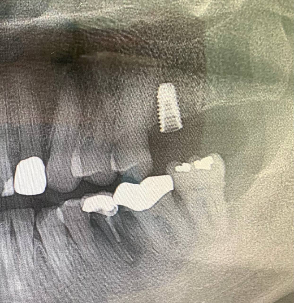
This PA is from a new patient to our practice. They have had quite a bit of work done in their country of origin and there are many issues including poor periodontal health and many failing restorations. The implant supported bridge seen here exhibits severe gingival inflammation and it is impossible to pass a floss threader under any portion. Also there appears to be residual cement under the prosthesis. Given the unfortunate position of the implants (which appear to be well integrated), I don’t know if removing this and replacing it with a screw retained prosthesis with better gingival contours will help much given the mesial and distal cantilevers. I am also not sure what type of implants these are? Suggestions?

8 Comments on Implant supported bridge with gingival inflammation: suggestions?
New comments are currently closed for this post.
Alex S. Abernathy
12/18/2017
You might consider removing the prosthesis and recontouring and polishing the tissue side . One could also currette the tissue while the prosthesis is out. The problem with this approach is "you break it you own it" especially if the patient has no perception of a problem.
pradeep kumar.dr
12/19/2017
please dont use word gingiva for implants.
its peri implant mucosa
Dr. TK
12/19/2017
This case is precisely why we should screw retain prosthetics whenever possible.
The retained cement needs to be taken care of. It looks like bits are floating around, but if you can't floss under it then we can almost guarantee that some cement was left behind at delivery. There is certainly enough evidence that cement is retained that a reasonable patient could be convinced to address the issue.
Here is what I would do First: a long conversation regarding condition, risks and possible outcomes. Second: send your x-ray to Attachments International for implant identification and make sure you understand the system. Third (treatment): make two screw access holes through the occlusal of this cemented bridge. Use your best estimate where the abutment openings are, drill straight through the porcelain and expand the hole until you find the screw access holes in the abutments. After removing both screws, the bridge and abutments will come out in one piece. Don't attempt to separate the abutments from the bridge, treat it as if it is a routine three unit screw retained bridge. Now you can reshape and polish the tissue side of the bridge, clean up cement and then deliver it (using new screws) as if it were a screw retained bridge.
There are potential issues (porcelain fractures, misjudging location of screw holes, previous dentist did not block out abutment opening so cement covers screw hole); however, the risk of no treatment are pretty severe in this case, so the treatment is certainly justified.
Dr. Rayment
12/19/2017
Thanks TK. That sounds like a good plan. I had considered trying to replace the entire prosthesis to minimize the cantilever effect but don't see how that would be possible without repositioning the implants. Hopefully we can get this "peri-implant tissue" in better condition.
denis cunneen
12/19/2017
DrTK has excellent solution
Recontouring the bridge out of the mouth to allow easy accessibility,The distal and medial extensions are not going to cause any issue ,
Dok
12/20/2017
First recontour the prosthesis gingival embrasure spaces to allow for cleansability ( small/thin flame shaped diamonds and finishing burs ) then open curettage, clean/disinfect the implant/abutment surfaces thoroughly and close the tissue.
Dok
12/20/2017
ALMOST FORGOT THE MOST IMPORTANT PART. AFTER TREATMENT, INSTRUCT THE PATIENT TO CLEAN BELOW THE PROSTHESIS USING AN IRRIGATING FLOSSING BRUSH. IRRIGANT SHOULD BE MIXTURE OF LISTERINE ( OR THE LIKE ) AND WATER IN THE RESERVOIR WELL, DONE TWICE DAILY. WHAT IS AN IRRIGATING FLOSSING BRUSH YOU ASK ?? E-MAIL ME AND I WILL SHOW YOU AN IMAGE OF ONE.
greg steiner
12/28/2017
Your periapical radiograph gives beautiful detail. It shows the residual graft material surrounded by radioleucencies. These radioleucenies have been found to be areas of hypervascularity and I believe this contributes to the slow exfoliation of the graft material. There is significant exfoliation of graft material at the crest between the two implants and throughout the gingiva. The gingiva in this radiograph is filled with exfoliating graft material.
Changing the prosthesis will not effect the long term failure of the bone supporting these implants. I saw a case like this last week and I advised that the patient to leave the bridge and implants in place and do something about it when it becomes symptomatic.


