Straumann Implant failed after 7 years without any signs or symptoms?

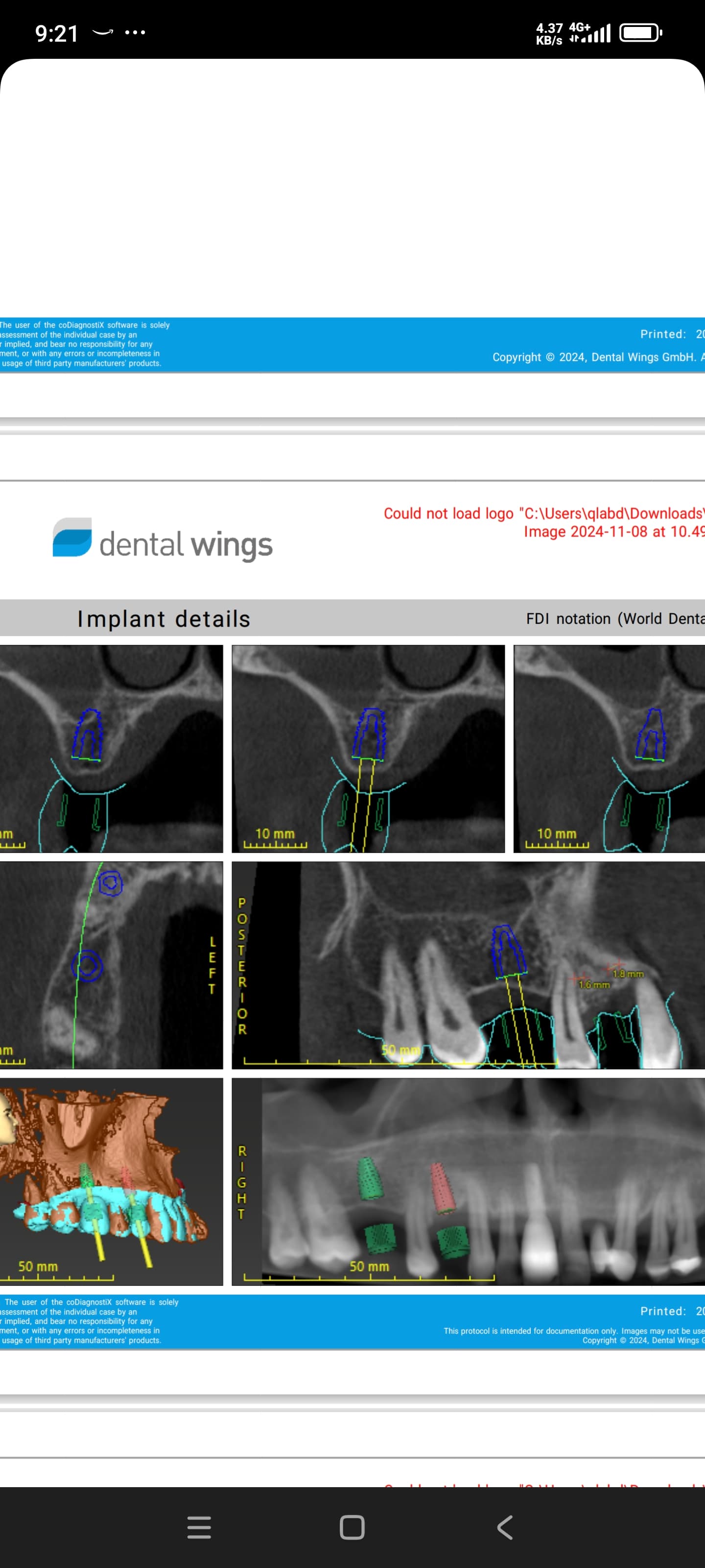
I installed a Straumann implant in #18 site [mandibular left second molar; 37] in 2005. Â It osseointegrated uneventfully and was restored by the referring doctor 6 months post-op. Â For the last 7 years there were no apparent problems. Â But 2 weeks ago, the implant became painful to chewing, loosened and actually came out. Â The crown was screw retained so there was no problem with cement extrusion. Â The implant was short. Â The only thing out of the normal that I noticed was that the opposing molar was in cross bite. Â Could occlussal trauma have caused this? Â Any ideas on causation or why there were no symptoms until just prior to loss of the implant?

(click image to enlarge)

![]David-implant-18](https://osseonews.nyc3.cdn.digitaloceanspaces.com/wp-content/uploads/2012/08/David-implant-18.jpg)

34 Comments on Straumann Implant failed after 7 years without any signs or symptoms?

New comments are currently closed for this post.
CRS
8/21/2012
I've seen this before, when an immediate implant is placed, you're stuck using either mesial or distal root so the crown is off center with almost a cantilever. Straumann inplants are very unforgiving due to the thread pattern so I only place them in solid well healed bone, grafted is okay. It was occlusal load that caused this to fail. just do a revision, remove it graft it with cadaver bone and prp or pgrf. Let it heal and place a self taping implant.
CRS
8/21/2012
Almost forgot use a surgical guide and make sure that the implant is centered and line up with the upper tooth
CRS
8/21/2012
Actualy is that a straumann?
CRS
8/21/2012
I'm confused did you place another implant on the same spot? A nobel active? When this is restored the new crown will have the same off center problem.
BTH
8/21/2012
Not a Straumann implant, but I bet it is occlusion....
Don Rothenberg
8/21/2012
Don't you think that occlusion after 7 years is a bit strange...do you have any x-rays of years 1-7 ? It would seem that if it was a occlusal problem it would have showed up much earlier. I am interested to see other ideas.
DR S
8/21/2012
Hi I think the first 4 pics are of the straumann implant, and the rest seem to be the revised placement with a new implant more mesial to the initial failed implant. Just a guess but the static occlusion might be stable, but could there be an element of parafunctual activity putting undue pressure in lateral movement? Might be interesting to ask if any thing has changed in the months coming to the loss of the implant.
Carlos Boudet, DDS
8/21/2012
A short implant in the second molar area receives the greatest masticatory forces, and has a higher chance of failure by occlusal overload than other areas of the mouth. There may not have been symptoms, but there will be signs, usually a slight radioluscence around the implant (sometimes not so slight), softer or dull percussion, etc... What size implant? Immediate placement? I would have waited for healing before placing the implant in that extraction site. Good luck!
Michel P. Jazzar
8/21/2012
The straumann implant has like a smiling face when you look on the Xray. I guess it is a bruxomania?
Vipul G Shukla
8/21/2012
I don't actually see a full cross-bite, rather a very pronounced palatal cusp on the upper 2nd molar, but the initial implant looked well-centred. The cantilever effect may be to blame for this failure, if predominant occlusion was in the mesial aspect of the crown, but then how do explain the success of All-on-4 concept? Was the implant body actually fractured? Anyways, unless the patient has uncontrolled diabetes or very advanced chronic peri-implantitis, this case does not make sense.
mjohnson dds, ms
8/21/2012
I agree with an earlier presenter. If it was occlusal overload why didn't it show up earlier? Why do we always have to blame something. Mother nature doesn't always cooperate with us. It could have just been been bad luck or poor protoplasm. I agree, the second molar site certainly takes the greatest stress so I am selective as to whether I replace them or not. I always question occlusal overload as an excuse. Why, if we leave a crown high, the opposing tooth gets mobile and hurts. Why doesn't the opposing tooth have symptoms if it's in hyperocclusion with an "ankylosed" tooth (implant). Why does the implant deintegrate rather than making the opposing tooth mobile? I know there are a lot of questions but we don't have all the answers and I think "occlusion" or "cement" gets blamed for more than their fair share of failures. Sometimes it's OK to say "I don't know why it failed"
Greg Steiner
8/21/2012
This was not an implant failure this is a graft failure. The site was grafted with a nonresorbable material that most likely produced sclerotic bone. This implant did not just fail overnight. The process started the day the implant was placed into occlusion. Because the "bone" the implant was placed into has no vascularity and no regenerative cells it is like putting an implant in concrete. The failure process in like breaking concrete with a sledge hammer. Each time you hit it a minor crack appears and after many small cracks(microfractures) the concrete finally breaks apart. Everyone who uses nonresorbable bone grafts like allografts or xenografts that produce sclerotic bone will see many failures like this one in the future. If anyone wants to see the histologic process of sclerotic bone failure email me and I will pass it one to you. Greg Steiner Steiner Laboratories
alupigus
8/21/2012
Even a short implant shouldn´t get disintegrated if perfectly integrated for 7 yrs- btw, how did the x-rays in between look like? However, I can think about some considerations which could lead to such a situation, where STRESS seems to be the prevailing issue here as: 1) straumann short implant has just very few threads ( up to three!!! Try counting them on your xray and compare to other manufacturers specialized on short implants! http://www.denfo.de/Implantate_3.JPG AND http://ars.els-cdn.com/content/image/1-s2.0-S1010518207001229-gr4.jpg ) which in cases of 2) mechanical overload "help" then as severe pressure peaks causing osteolysis to the surrounding bone making the implant fail (which is btw not mandatory to be diagnosed on check-up x-rays). 3) Parafunction could add to it. 4) Your posted pictures show that there must have been at least one cantilever (mesial) when looking at the crown on the picture taken after the failure. If there is a cross bite, only from a cat scan you can tell how the direction of the bite force was( where the direction of the force is much more important than the amount). If it was strictly axial it shouldn´t be the cause of failure. If it´s off axis there you might have your second cantilever (vertical cantilevers causing shear load on the bone by an implant with just 3 threads!!!). In my view the amount of time (7yrs) speaks quite clearly for the critical impact of things like: -shear loads -vertical cantilevers -mechanical overload -direction of the forces on a presumed perfectly osseointegrated implant making it fail in the end, because if we think about the most well known Stress Theorem where: STRESS = FORCE / AREA then, if the applied forces are in balance with the area of the well integrated implant, nothing bad should ever happen. But who can balance that perfectly out incl. parafunction, position of the implant, inclination, prosthetic work, excursions, etc,etc.? Therefore, I think that single 6-8mm Implants for a molar (where god put in 2 roots in stead of just one compared to the front) might be sometimes under-engineered, where we would be far better off to overengineer. It´s just my take on the issue. Each and everyone may adress it differently... Hope this was of help.
Rand
8/21/2012
Over the last twenty years I have seen 4 cases like this. They each had one common factor. Each was a man. Each had broken a tooth followed by successfull endodontic treatment post and core and crown, followed some years later by root fracture and extraction. One of the four cases, the implant did not loose osseointegration, it broke in half. Patients who are hard on their occlusion will cause wear. An implant that had very kind occlusion 7 years ago in such patients would be in hyper-occlusion and be heading for failure now. Moral of this scenario: Perfect implant placement needed to optimize occlusal forces followed by occlsal force monitoring each year.
Ítalo José Vitorino Net
8/21/2012
I agree with Rand, the fact that seven years has passed, makes me think about overload over the time, perhaps the other teeth slowly has been worn and the porcelain did not, so the contact between the implant prothesis and his opponent tooth started the problem! The rest is history! Maybe, with more precise occlusal control and adjustement the end could be better, like prognostics
Greg Steiner
8/21/2012
The replacement implant was placed in the old graft material which caused the failure in the first place and in addition the graft material that was added appears to be similar graft material which will never integrate to the new implant surface. If the replacement implant integrates I give it a year at most. When that one fails remove all of the nonresorbable graft material and graft with a resorbable graft material and let it become viable bone before placing your next implant. Greg Steiner Steiner Laboratories
.
8/24/2012
Why should anybody graft a nonresorbable material in the human bone? That makes no sense.
Perioperry
8/21/2012
Recently had a case almost the same as this. Straumann implants in #18 and #19 sites, crowns not splinted together. The #18 implant "dis-integrated" after 9 years and came out easily during procedure to make new crowns on the implants. I think it was occlusal stress. I think splinting adjacent implant units is almost always advisable, but this case was not done this way.
sb oms
8/21/2012
to greg steiner- what are you talking about- graft failure? what graft? there is no mention of a graft in the original placement. Did i miss something? i've read hundreds of your comments, they are all pretty much the same - you are obviously a fan of resorbable graft material - so am I. But what does that have to do with this case? i've seen some second molar implants fail in this exact manner, and my bet is overload. how to treat, how to avoid - pending...........
Greg Steiner
8/22/2012
sb oms Please take a look at the radiograph and the graft is clearly outlined. If you are in doubt ask the poster what graft material was used and I will be able to explain more accurately how this failed. Few of the posts on this case mention anything other than occlusion or implant design and few are discussing the material that is supporting the implant namely the bone. Is there any question that what failed here is the bone that supports the implant? Because the bone failed the discussion should start with questioning if there was something wrong with the bone before looking for secondary causes. There may be contributing factors but this is a graft failure. Greg Steiner Steiner Laboratories.
sb oms
8/21/2012
i just don't think you can explain this with a product discussion or brand comparison.
naswe
8/22/2012
there are so many factors could ve caused this failure - short implant in the area of 2nd molar where heavy force of mastication takes place -cantilever effect of the crown -crown placed in cross bite where shear force and off access pressure effect takes place -over loading -general factors such as smoking,diabetes,bad oral hygiene etc
rsdds
8/22/2012
in my opinion implant failure in the lower 2nd molar areas are very common that's why i choose no to restore this area unless implants in this area are splinted to an implant in the first lower molar site, or the patient really wants it and is fully aware of the possible complications..
dro
8/22/2012
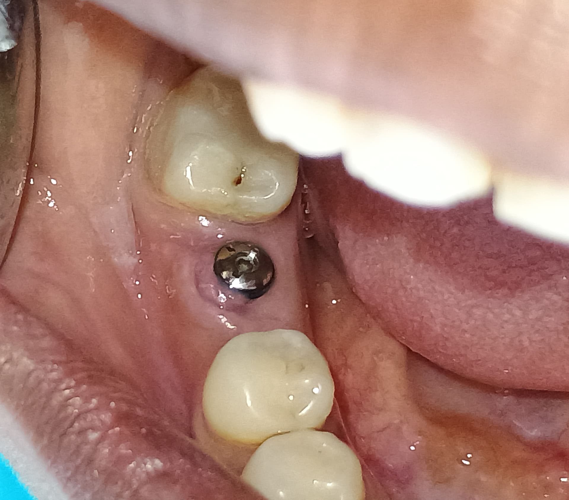
If you look at the picture where the implant has been removed you can see dense bone circumferentially around the prior implant site. This indicates to me that it has been subjected to substantial occlusal forces for some time. While we intend for function to occur immediately after contact between natural teeth, time, wear, bruxism, supereruption and degeneration within the TMJ can and will alter functional and parafunctional relationships between teeth. We can do the best we can to adjust for these situations but it is impossible to do so in all circumstances. In other words, defecation happens. I do feel it will likely recur in this patient given enough time if the occlusal load cannot be controlled.Good luck!
DENIS CUNNEEN
8/22/2012
The occlusal stress may well have been rotational, creating a torquing moment. The ITI implants can just unwind.The large cantilever style crown and the reliance on the distal ossification of the Bioss? material my have contributed.
Dr. Joe Jaws
8/22/2012
Dear fellows: what a discussion.... You learned that an implant is a device that replaces the missing root of a determined tooth.So if the lower molar carries 4 roots fused into 2, common sense tells us we should place 2 implants to replace such roots.Use your brains and do not load 2 impacting forces over 1 support, but rather 2 over 2. End of problem. In this case, the implant started to fail,the day you applied the final torque.
PhD. BOJI SAAD
8/22/2012
so i will not agree with the cause of occlusion ...i want to ask if the patient have systemic disease like diabetes or osteoporosis and is male or female ? does she or he under some medication the last years like bisphosphonate therapy ?,which can cause osteolysis very easily
Dr. Gerald Rudick
8/22/2012
In my opinion, the photos are not placed in the correct chronological order so it is difficult to give a clear opinion. Dr. Greg Steiner brings up a very excellent point, in that waiting 4-6 months after a grafting procedure, when attempting to place an implant....it goes in very solid...and you think you have a great result.......but aside from autogenous bone,the commercially available synthetic bone materials, as well cadaver bone is not living and you may be drilling into concrete that has no blood supply and very little osteoblasts or osteotcytes.......the initial stabilzation is mechanical and not biological. Excessive forces can cause pressure necrosis and cause live bone to "give up" and detach itself from the implant surface. The honest and scientifically knowledgeble graft purveyors will tell you that it can take years for a grafted bone site to remodel itself and become natural bone........( some materials never resorb) very frustrating to see a beautifully engineered case fail. Dr. Gerald Rudick Montreal, Canada
Greg Steiner
8/22/2012
Dr. Rudick Thanks for the backup. Your description was better than mine. Greg Steiner Steiner Laboratories
peter Fairbairn
8/23/2012
Greg I also agree with your rational which was backed with a long discussion with a father of ITI about the presence of Osteocytes in grafted sites denoting living bone . As loss it could a combination of many factors as this can happen , and has happened to me in the lower 7 area . Strangely a patient came in to see me with a handful of short tissue level implants that had fallen out , but looking at the positioning of the remaining ones I could see why. Regards Peter
Richard Hughes, DDS, FAAI
8/23/2012
Consider longer implants.
Jennifer Watters, DDS
8/28/2012
I agree with M Johnson, DDS. Sometimes we don't know the cause of failure. There may be other "patient-related causes" i.e., systemic issues at play also. I have witnessed the loss of a "2nd molar implant" when placed within the natural dentition as the posterior-most tooth. I haven't seen this with a "1st molar implant" with a natural tooth posterior to it. I don't think it can be only about occlusal factors so many years later..... I also had this happen about 7 years after successful placement and function in a non-smoker who did have a heavy occlusion, but all of the other implants in his posterior mandible were fine even the contra-lateral side. Go figure?
CRS
9/4/2012
I was thinking about this, was there any peri-implantitis prior to the avulsion of the implant? About 7-8 years after placement is when we start to see this.Implant "grafting" is really space maintenance while allowing the body to regenerate new bone, it is not like placing an implant in "concrete".While some xenografts will become incorporated within the bone matrix, there are no live cells being transplanted. The original implant integrated, but probably over time (cantilever) the implant was lost due to occlusal wear.I would have regrafted the area and waited then placed a new implant in an ideal occlusal position not allowing the available bone to dictate placement. And I would have the restoring doctor evaluate the occlusion prior to restoring. A lot can happen in 7 tears and as dentists we tend to blame ourselves for "failures" (vs a 7year success) and performing a "revision" like the orthopods do.
Huda Albather DDS, MDS, M
9/4/2012
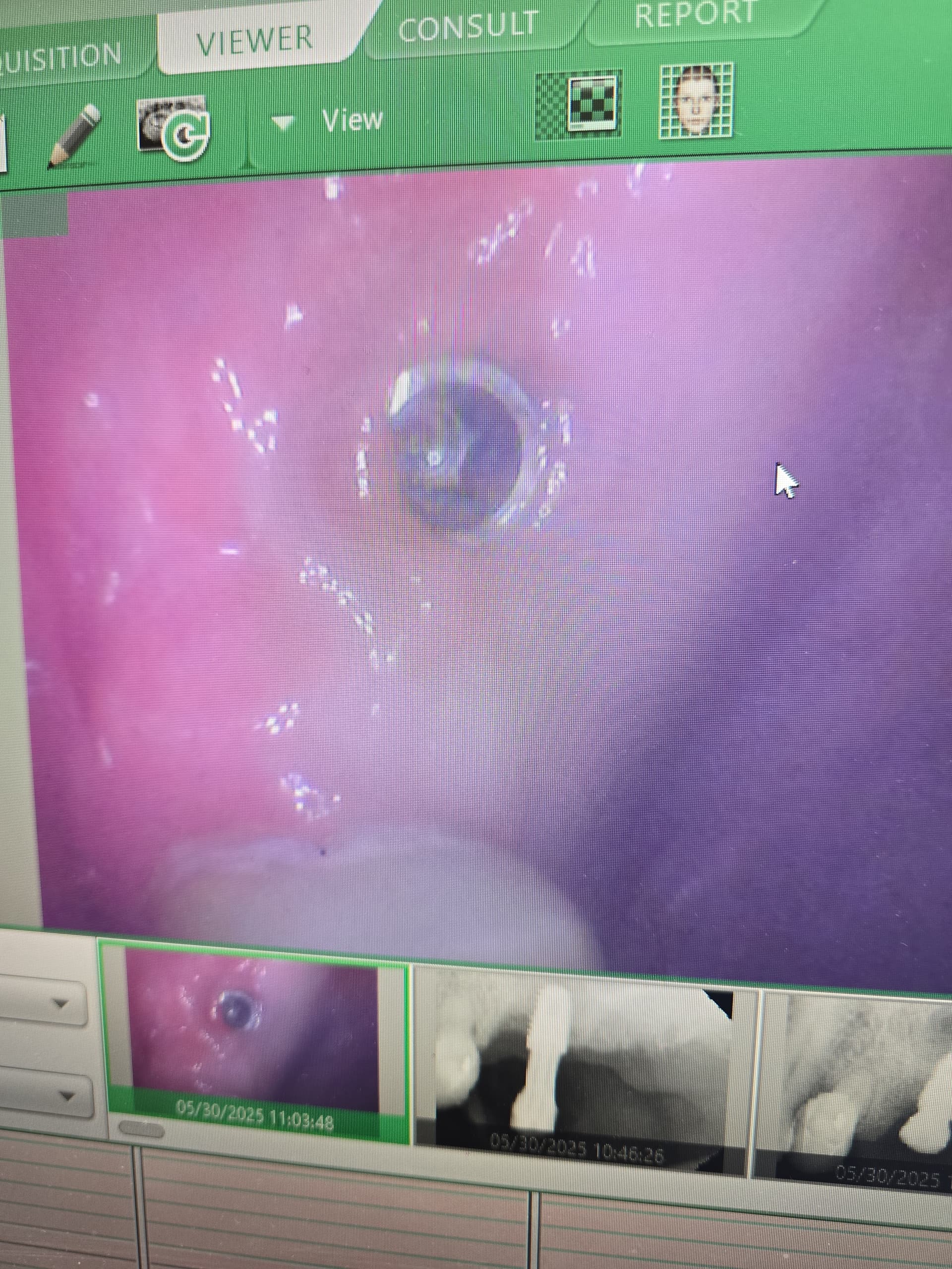
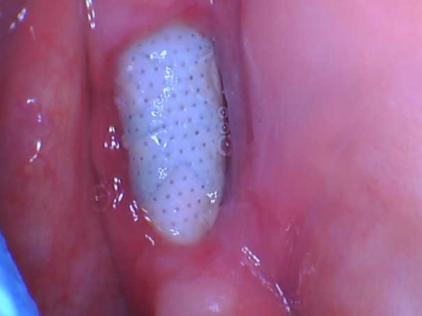
Thank you all for your responses. The patient is a 50 year old non-smoker healthy male. The pictures are in the correct chronological order as follow: first picture is from 2005 before sending the patient to his dentist for final restoration and the first x-ray is also from 2005 few months after her had the implant restoration. The second x-ray is from 8/2012 when he showed up with the implant in his hand (one piece no fracture) just fallen out and that he did not experience any swelling, infection or pain, only slight discomfort 2 weeks prior. The rest of the photos are also recent which shows the implant socket with granulation tissue, and the position of the new implant (Noble bone level) with more mesial placement. The old implant was Straumann tissue level SP size 8 x 4.8 mm WN and the new implant is Noble active internal bone level size 10 x 5 mm. I have a CT scan in my office which I did not have in 2005; however, on the scan the position of the old implant was also centered with bone all around!!

Featured Products

OsteoGen Bone Grafting Plug
Combines bone graft with a collagen plug to yield the easiest and most affordable way to clinically deliver bone graft for socket preservation.
CevOss Bovine Bone Graft
Make the switch to a better xenograft! High volume of interconnected pores promotes new bone. Substantially equivalent to BioOss and NuOss.