Loss of gum due to laser: does this case need a gingival graft?
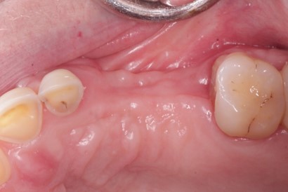
I have exposed an implant with a laser in an anterior case where keratinised gingiva seemed abundant. When I placed the healing collar, the buccal gingiva was very thin and now it is gone. I had put the cover screw on and the patient is under observation. Do you think now that the healing screw is off the tissue will grow back or does this case need a gingival graft?
 anterior
13 Comments on Loss of gum due to laser: does this case need a gingival graft?
New comments are currently closed for this post.
CRS
4/16/2013
The mucosa was most likely lost due to no bone on the buccal, since the implant was placed too deep in order to compensate. The implant is not in the dental arch. I would suggest removing the implant, grafting the lost bone and closing the mucosa. Regardless of the thin bio type and triangular shaped teeth which are caution signs in themselves, that much mucosa cannot be grafted without bone under it to support it. You also could put the implant to sleep and try to graft over it but I'm not sure that will work. I feel that this is just really poor understanding of implants in the esthetic area. Now there is a nice cleft. It is very important to respect the buccal plate. I'm sorry to be so blunt, but I can't fix that clinically. The implant sites have to be developed prior to implant placement it has nothing to do with the laser, it just happened sooner, since the blood supply was compromised in the flap. You could try to place a flipper to cover the defect in the meantime, hopefully the adjacent teeth will not develop bone loss. This is not a restorable implant. Get some help with this case. Thanks for reading.
Sam Jain
4/18/2013
Can u put a impression coping on the implant and post the picture
If implant is infact placed facial, no tissue evaporation by laser should have been
done....all the tissue needs to be preserved.
It is a big problem....pl post pictures of the healing tissue
Sam Jain, DMD
Center for Implant Dentistry
Fremont, CA
Sam Jain
4/18/2013
Pl take a picture with impression coping and post it. As general rule, never evaporate facial tissue on anterior teeth....always preserve.
Pl post pictures of the healing of the soft tissue with the cover screw.
Sam Jain DMD
Center for Implant Dentistry
Fremont CA
ttmillerjr
4/21/2013
Thin biotype CRS?? This is classic thick. As for this case, the patient has diastemas behind his laterals, is he missing his canines. It looks like he is in group function with his laterals dragging across his lower anteriors, plus his remaining central looks as wide as it is long. I think one could make an argument for rebuilding his occlusion. I'd consider a little crown lengthening and bridges; one from 5-6-7-8-9 and one from 10-11-12. This way you could establish a more functional occlusion, "replace" his missing teeth and get a great esthetic result.
Not every case is an implant case. I agree with CRS that this is poor planning. It's easy to think that it's only one tooth so no need to tx plan, but this is a good example of why planning is needed. This case demonstrates ignorance of even the most basic knowledge of implant surgery.
Richard Hughes, DDS, FAAI
4/21/2013
Dr ttmiller, I agree with you 100%. This is a classic thick biotype and a classic work wp with mounted cast, wax-up, occlusal analysis is the way to start. Classic crown and bridge and crown lengthening is a wiser treatment plan. I agree, Implants are not always the treatment of choice. Maxillary C & B from first bi to first bi will yield superior esthetic results.
CRS
4/22/2013
Sorry I was hasty in looking at the thick gingiva and shape of the crowns, but this implant was placed way too deep which makes me suspicious that there is no bone on the labial and this is a pretty good example of what not to do with a laser. I think what triggered me is that the poster thinks this will heal and somehow thinks this is just from using a laser, look at the placement. I definitely agree that this treatment focused solely on placing an implant without regard for the occlusion or the diastemas. A lot of corners cut! It seems it was a rush just to place an implant. Thanks for reading crown and bridge gurus!
Drbddsinc
4/23/2013
Lets assume the patient doesn't want implant removed. This case can be "salvaged" with a custom abutment and the lab can use some pink porcelain. This case is far from ideal now anyways. Would further surgeries improve things for certain?
mahendra azad
4/24/2013
I agree with CSR. This has nothing to do with Laser.
Dr Bob
4/24/2013
Drbddsinc
Yes, removal of the implant and grafting to regain bone and other missing tissue would improve the physical condition. Restoration with a custom abutment and a lot of pink porcelin might be acceptable to the patient as a short term solution, but is this a predictable long term fix? The patient must advised to what future out comes and what future costs to correct these problems are likely to be and then and be involved in the decisions as to how to proceed. It would be deceptive (even if unintentionable) to restore with a custom abutment with out advising the patient of how with time the aesthetics could change. I know that from the way that you wrote "salvaged" you did not suggest a deception, but the presenting doctor is new in the field of implants and must be made aware that a custom abutment could provide a temporary solution that could result in a future nightmare.
Sam Jain DMD
4/26/2013
I think it would be better to bury the implant instead of removing the implant. Put cover screw, de epithelial the soft tissue. Take molt and scrape off epith from max tub crestal tissue and then take out the entire thickness of tissue and put it like a wedge in the cleft.
And then crown and bridge at NO CHARGE. Inform px of YOUR mistake/irresponsible behavior.
And life moves on.
Sam Jain, DMD
Fremont implant dentist
CRS
4/27/2013
That sounds like a good idea do you have a reference or photo of technique ? Could bone be grafted over also? Please advise.
Gregg Weinstein
5/3/2013
Take the implant out asap before the apical portion integrates. The loss of tissue is from lack of underlying bone crestally (source of vascularization)... just about impossible to graft bone on the coronal portion since the surrounding bone on the adjacent buccal plates is probably the same thickness or thinner. Remember, you will only get the thickness of the surrounding plate long term with a graft. Remove the fixture and graft with a coronally positioned flap. you could use a trephine graft from the maxillary tuberosity if possible? After healing you could place a fixture with a palatal incision and apically repositioned flap to possible regain that nice kertinized tissue)
CRS
5/22/2013
I have a similar case which I hope to post the patient came in for a second opinion very deep placement and I have her consulting with my prostodontist as my wing man. Do we patch it and let it fail or do I go in guns ablazing and fix now 12 weeks after placement by a very interesting "colleague". It will be a challenge for me to post since I am not good with the computer. I really value the judgement of my dental posters out there who are in the implant trenches! Stay tuned! This might turn into a CRS roast!


