Socket Grafting and Ridge Preservation using Bond Apatite

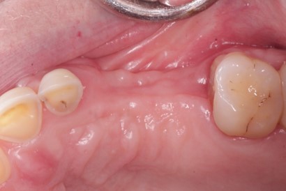
In this case, by David Baranes D.M.D, the two inferiors hopeless molars 37,38 were removed. The socket and the ridge were preserved by augmenting the area with Bond apatite bone graft cement. After teeth removal and complete debridement of the site, the application of the cement was made according to the protocol in 3 consecutive steps: place,press, and close. The cement was ejected directly into the grafted site from “all-in-one” syringe and then press firmly with a dry gauze for 3 seconds, followed by primary soft tissue closure directly above the cement. No membrane was needed. 12 weeks post-op, at reentry bone was formed and the 3D ridge volume was preserved. At this stage implant placement took place. Please share any questions/comments.


> Editor’s Note: Bond Apatite is a grafting product that combines biphasic calcium sulfate with a formula of hydroxyapatite granules in a pre-filled syringe to create a self-setting cement for bone graft procedures. Watch Introductory Bond Apatite Videos or Learn More.

10 Comments on Socket Grafting and Ridge Preservation using Bond Apatite

New comments are currently closed for this post.
Sean R
6/30/2017
What a nice case. The implant seems well positioned with lots of bone. Three months seems a little quick for placement but seems much more common today. How long do you like to wait with grafting versus without? Of course this may change based on location and bone quality?
Dr .Amos Yahav
6/30/2017
in this case due to the graft nature and its ability transform into the patient own bone 3 months are a very reasonable time to place or load the implant . when using a graft that integrate with the bone and du not replaced by the new formed bone than you should wait until maturation 5-6 months at least if you like to work on the safe side
Kaz Zymantas
7/2/2017
What do you do if you cannot get primary closure? Thanks
Dr. Amos Yahav
7/2/2017
In socket grafting cases, you don't need to reflect the flap. However, you need to protect the graft during the healing. You can do so by using any barrier that can last for an average of 10 day for example any collagen dressing (not a Gell temp ) can be used which should be sutured and attached to the surrounding soft tissue ,followed by a criss-cross suture above it. In other cases, closing of the flap is not as we are used to doing when we are using granules and membranes, where we need to have tension free and primary closure.With this type of cement, we want that the flap to be with slight tension and primarily closed. Also, if you have exposure during flap closure of 2 mm it is not an issue. But, not more than that, in order not to lose volume. You can see the technique in this video on Lateral Augmentation with Bond Apatite.
Peter Fairbairn
7/4/2017
Yes Amos , this is the most efficient route to help the Host to regenerated bone ... nice case. Peter
Dr. Amos Yahav
7/4/2017
Thank you Peter indeed we strive to regenerate and not just to integrate .
Michael
12/14/2017
Bond Apatite has HA in its composition (33% or so). Any concern with the HA not being fully resorbed? I heard that some HA with high crystallinity cannot be resorbed by the body.
Dr Yahav Amos
12/15/2017
most of the existing bone grafts are not resorb they integrate with the new formed bone therefore the amount of vital bone is more or less about 20-25% and not more than that .even allograft that many clinicians tend to believe that it turn into the patient own bone .which is definitely not the case . it just integrate with bone that is built between the allograft particles, and the proof is that you will never find any osteocyte within the Allograft particles,which shows that there are sterile sequestrum but not vital bone .they integrate very nicely but not become bone ,you can see it as well during the radiographic follow up it is always radio opaque during healing which shows that there is no any transformation .the same of course is with xenografts and the majority of the synthetic material. by using the Bond Apatite on the contrary the Biphasic calcium sulphate matrix is transform completely and simultaneously into the patient own bone therefore in 3 months you will have 3 times more vital bone than any of the existing materials and the remaining HA particles 33 % due to their particles size differences and shapes resorb as well the small to medium sizes in 3-6 month and the rests wich are less than 10% remain a bit longer .so if you compare it to the poor vital bone quantity of most of the others there is doubt that by using this type of graft your vital bone quantity is the highest one and much more than the others .
Michael
12/15/2017
Dr. Yahav Amos, do I understand correctly that Bond Apatite is 100% resorbed and replaced by vital host bone in less than a year, regardless of graft size?
Dr. Amos Yahav
12/15/2017
3 D bond in 3 months is completely 100% transfer into the patient own bone , however since it is a short term space maintainer, its indication by itself are limited to socket grafting procedures . on the other hand Bond Apatite as a composite graft its indication are not limited and most of it will become the patient own bone after one year, as i mention earlier the large particles of HA which are less than 10% their resorption might take longer . depend on the defect size ,morphology ,patient etc .

Featured Products

OsteoGen Bone Grafting Plug
Combines bone graft with a collagen plug to yield the easiest and most affordable way to clinically deliver bone graft for socket preservation.
CevOss Bovine Bone Graft
Make the switch to a better xenograft! High volume of interconnected pores promotes new bone. Substantially equivalent to BioOss and NuOss.